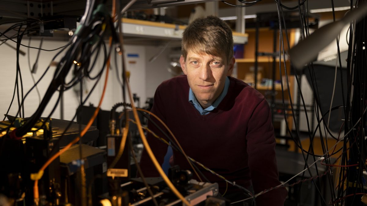
Adam Cohen.
Niles Singer/Harvard Staff Photographer
Health
Tracking precisely how learning, memories are formed
Groundbreaking new technique may offer insights for new therapies to treat disorders like dementia
A team of Harvard researchers have unveiled a way to map the molecular underpinnings of how learning and memories are formed, a groundbreaking new technique expected to offer insights that may pave the way for new treatments for neurological disorders such as dementia.
“This technique provides a lens into the synaptic architecture of memory, something previously unattainable in such detail,” said Adam Cohen, professor of chemistry and chemical biology and of physics and senior co-author of the research paper, published in Nature Neuroscience.
Memory resides within a dense network of billions of neurons within the brain. We rely on synaptic plasticity — the strengthening and modulation of connections between these neurons — to facilitate learning and memory.
Synapses, or the junctions where neurons communicate, lay the groundwork for every memory we form, from a childhood melody to a loved one’s face to what we ate for breakfast.
In their new paper, the team detailed their new technique, dubbed Extracellular Protein Surface Labeling in Neurons (EPSILON), which focuses on mapping the proteins vital for the transmission of signals across synaptic connections in the brain.
Utilizing sequential labeling with specialized dyes, EPSILON enabled the researchers to monitor these proteins’ movements at high resolutions.
Credit: Pojeong Park
These specific proteins are called AMPARs and are considered key players in synaptic plasticity, the process that allows the brain to adapt and reorganize itself in response to new information.
Utilizing sequential labeling with specialized dyes, EPSILON enabled the researchers to monitor these proteins’ movements at high resolutions. Traditionally, understanding such detailed microscopic phenomena has required more invasive methods. Using EPSILON to observe AMPARs’ behavior in neurons represents a significant scientific advance.
This work was undertaken by several members of Cohen’s lab, including Harvard Griffin GSAS student Doyeon Kim and postdoctoral scholars Pojeong Park, Xiuyuan Li, J. David Wong-Campos, He Tian, and Eric M. Moult, as well as scientists from the Howard Hughes Medical Institute.
A combination of fluorescent labeling and cutting-edge microscopy allowed the researchers to illuminate synaptic behavior at unprecedented resolution. The technique’s precision was akin to shining a spotlight on some of the brain’s most intricate functions, allowing the team to monitor the synaptic interactions critical for learning.
As synaptic changes of specific memories came into view with greater clarity, patterns started to reveal rules governing how the brain decides which synapses to make stronger or weaker when storing a memory.
Prior research into synaptic processes often lacked such granularity, making EPSILON’s insights particularly valuable for future explorations into diseases like Alzheimer’s, marked by synaptic dysfunction that results in memory and learning impairment.
As synaptic changes of specific memories came into view with greater clarity, patterns started to reveal rules governing how the brain decides which synapses to make stronger or weaker when storing a memory.
“Our most important breakthrough is our method that can map the past history of the synthetic plasticity in the living brain,” Kim said. “We can look at the history of the synaptic plasticity, studying where and how much of the synaptic potentiation has happened during a defined time window during the memory formation.
“By mapping the synaptic plasticity over time at multiple time points, we can truly map the dynamics of the synapses,” Kim added. “We’ll also be able to apply this to different kinds of memories that have different patterns of synaptic plasticity.”
The technique’s first application has already yielded intriguing results. By applying EPSILON to study mice undergoing contextual fear conditioning — a process that helps animals associate a neutral context with a fear-inducing stimulus — researchers were able to demonstrate a correlation between AMPARs and the expression of the immediate early gene product cFos, a signal that tells us when brain cells are active.
These findings suggest that AMPAR trafficking is closely linked to enduring memory traces, or engrams, within the brain, where specific neurons become activated following learning experiences.
Cohen credits the significant and sometimes unexpected role basic science can play in fueling progress with enabling their study to succeed.
“The HaloTag technology, which is used to label proteins, was based on a gene discovered in 1997 by a group of scientists in Ireland who were studying a soil bacterium, which had an unusual ability to break down pollutants,” Cohen said. “It’s a generations-long arc from the basic research characterizing the natural world to making discoveries that can make human health better. We really need to support the entire arc to make progress.”
Looking forward, Cohen is eager to see how EPSILON can be further applied to study numerous cognitive phenomena and potentially improve therapeutic strategies targeting memory impairments.
“We’ve already distributed the molecular tool to labs around the world who are now starting to use these tools to explore how synaptic strength is regulated in their favorite question and context,” he said.
This work was partially supported the National Institutes of Health.

